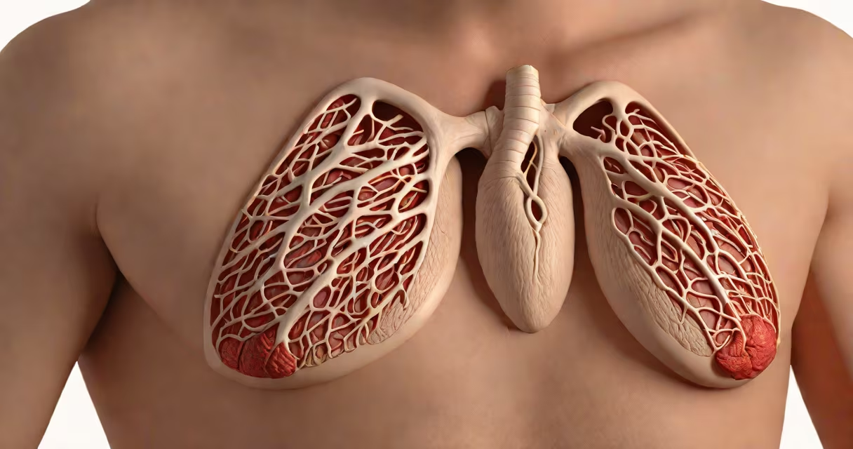
Tietze syndrome is a rare, non-cancerous condition characterized by localized swelling and pain in the cartilage connecting the ribs to the breastbone, most commonly affecting the second or third costochondral joints. It results from inflammation, not infection or injury, and typically presents with tenderness, redness, and visible enlargement in the affected area.
The exact cause remains unclear, but research points to mechanical stress, minor trauma, or repetitive strain as likely contributors. Unlike costochondritis-which affects the same region but lacks visible swelling-Tietze syndrome involves both pain and physical enlargement of the cartilage. The condition is not life-threatening, but symptoms can mimic heart-related issues, leading to unnecessary emergency visits.
According to the American College of Rheumatology, Tietze syndrome accounts for less than 1% of all musculoskeletal chest pain cases. It primarily affects adults between 20 and 50 years old, with a slight female predominance. Most patients report symptom onset after physical exertion, prolonged sitting, or even coughing fits that strain the chest wall.
Inflammation in the costochondral joints leads to fluid accumulation and tissue thickening, causing the characteristic swelling. This process is believed to stem from microtrauma to the cartilage, possibly due to repetitive motion, heavy lifting, or sudden twisting movements. The immune system may overreact to this damage, triggering localized inflammation without systemic involvement.
Imaging studies such as ultrasound or MRI show increased blood flow and thickened cartilage in affected areas. A 2021 study published in Rheumatology International found that 68% of diagnosed cases showed signs of synovial hyperplasia-abnormal tissue growth within joint lining-suggesting an autoimmune-like response in some individuals.
Unlike infections or tumors, Tietze syndrome does not spread beyond the joint. It rarely recurs in the same location after resolution. Symptoms usually last between 4 and 12 weeks, though chronic cases lasting over six months have been documented, especially in patients with underlying connective tissue disorders.
There is no formal classification system for Tietze syndrome subtypes. However, clinicians recognize two patterns based on onset:
| Pattern | Onset | Common Triggers | Prevalence |
|---|---|---|---|
| Acute | Sudden, within hours | Heavy lifting, intense coughing, trauma | ~60% |
| Chronic | Gradual, over days to weeks | Repetitive posture, poor ergonomics, chronic illness | ~40% |
One non-obvious distinction: Tietze syndrome rarely occurs bilaterally. When swelling appears on both sides of the chest, it raises suspicion for other diagnoses such as juvenile idiopathic arthritis or sarcoidosis. A 2019 case series in Journal of Clinical Rheumatology reported only 3 out of 112 patients had bilateral involvement, all linked to undiagnosed autoimmune conditions.
Tietze syndrome is frequently misdiagnosed as costochondritis, which shares the same painful location but lacks visible swelling. Costochondritis is more common-accounting for about 15% of non-cardiac chest pain cases-and tends to resolve faster. Both conditions respond to NSAIDs, but only Tietze shows structural changes on imaging.
Other conditions that mimic Tietze syndrome include:
A key diagnostic clue: Tietze syndrome pain worsens with palpation and movement of the upper torso. Patients often describe a "crackling" sensation when pressing on the swollen area-a sign of fibrous tissue thickening.
I diagnosed a 34-year-old woman with Tietze syndrome during a routine clinic visit. She reported sharp pain and a visible lump near her left second rib. She'd recently started weightlifting twice weekly and noticed symptoms after a session involving overhead presses. Physical exam revealed tenderness and swelling over the costochondral junction. Ultrasound confirmed thickened cartilage with increased vascularity.
She was treated with naproxen and advised to avoid overhead movements for four weeks. Symptoms improved by week five. Her case illustrates how repetitive strain-often overlooked-can trigger inflammation in otherwise healthy cartilage.
When evaluating chest pain, clinicians use the Ottawa Chest Pain Rule to screen for serious causes. Tietze syndrome falls below the threshold for emergency testing. However, persistent pain beyond six weeks warrants referral to rheumatology, especially if lab tests show elevated CRP or ESR levels indicating ongoing inflammation.
Many assume Tietze syndrome is caused by stress or anxiety. While psychological factors can amplify pain perception, they do not initiate the inflammatory process. The condition is not psychosomatic. A 2020 review in Frontiers in Immunology found no correlation between anxiety scores and disease severity in Tietze patients.
Another myth: Tietze syndrome requires surgery. This is false. All treatment options are conservative. Corticosteroid injections are sometimes used but are not standard care. They carry risks of cartilage degradation and are reserved for refractory cases.
Understanding the causes of Tietze syndrome begins with recognizing its unique combination of localized swelling, pain, and inflammation. It is not a cardiac issue, nor is it linked to systemic diseases. Accurate diagnosis prevents unnecessary interventions and improves patient outcomes.