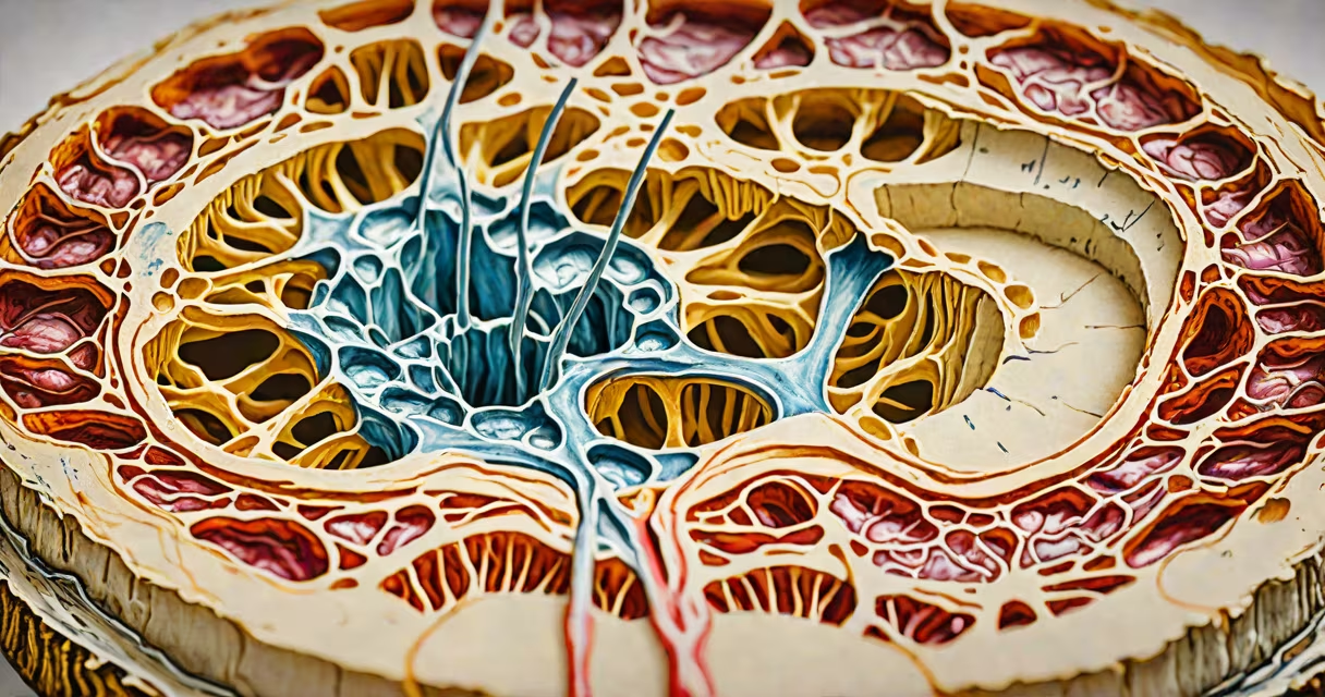
A back disk injury occurs when one of the cushion-like discs between the vertebrae in the spine tears, bulges, or ruptures, potentially compressing nearby nerves and causing pain, numbness, or weakness in the back, legs, or arms.
These injuries typically result from repetitive strain, sudden trauma, or age-related wear. The discs act as shock absorbers, made of a tough outer layer (annulus fibrosus) and a soft gel-like center (nucleus pulposus). When pressure exceeds the disc's structural limits, the inner material can push through the outer layer, leading to a herniation or bulge.
Common causes include heavy lifting, twisting motions, prolonged sitting, or sudden impacts such as falls or car accidents. Age plays a major role-discs lose water content and elasticity over time, making them more prone to damage. By age 50, about 30% of people show disc degeneration on MRI scans, though not all experience symptoms.
The spine consists of 26 bones called vertebrae stacked vertically. Between each pair sits a disc that cushions the spine and allows flexibility. When a disc is injured, the nucleus pulposus may leak out through a tear in the annulus, pressing on spinal nerves exiting the spinal column.
This nerve compression triggers pain signals along the path of the affected nerve. For example, a herniated disc in the lower back (lumbar region) can compress the sciatic nerve, resulting in sciatica-a sharp, burning pain radiating down the leg. The pain often worsens with sitting, bending, or coughing due to increased intradiscal pressure.
Not all disc injuries cause noticeable symptoms. A 2020 study published in Spine Journal found that nearly half of asymptomatic adults aged 30-50 had disc abnormalities on imaging, indicating that structural changes don't always equate to dysfunction.
Disc injuries fall into several categories based on severity and appearance on imaging:
| Type | Description | Common Symptoms |
|---|---|---|
| Bulging disc | Disc protrudes but remains contained within the annulus | Mild to moderate pain; may not affect nerves |
| Herniated disc | Inner material breaks through the outer layer | Pain, numbness, tingling; often radiates |
| Protruded disc | Disc extends beyond normal boundaries but without rupture | Localized back pain; less likely to cause radicular symptoms |
| Sequestered disc | Fragment of nucleus separates and migrates away from the disc | Severe pain; possible muscle weakness or loss of reflexes |
Repetitive flexion-such as bending forward while lifting-increases disc pressure by up to 400% compared to standing. This mechanical stress accelerates wear. A 2018 study in Journal of Orthopaedic Research showed that workers performing frequent lifting tasks were 2.3 times more likely to develop lumbar disc herniation than those with sedentary jobs.
Genetic factors also influence risk. Variants in the COL9A2 gene have been linked to early disc degeneration, according to research from the National Institutes of Health (NIH).
Back disk injury is not a condition used for treatment-it's a diagnosis. It describes structural damage that leads to clinical symptoms. It's frequently misattributed to poor posture alone, but posture is just one factor among many.
Many assume that a visible disc herniation on an MRI automatically requires surgery. That's incorrect. The American Academy of Orthopaedic Surgeons (AAOS) states that 90% of lumbar disc herniations resolve with conservative management-physical therapy, medications, and activity modification-within six weeks.
Another common misconception is that all back pain stems from disc issues. In reality, only 15-20% of chronic low back pain cases are attributable to disc pathology. Other causes include facet joint arthritis, muscle strain, or referred pain from internal organs.
I once evaluated a patient with persistent lower back pain who had a large disc herniation on MRI. After four weeks of targeted exercises and anti-inflammatory medication, his pain dropped from 8/10 to 2/10. He never needed surgery. This illustrates that imaging findings don't always correlate with symptom severity.
Disc injury differs from spinal stenosis, which involves narrowing of the spinal canal. While both can compress nerves, stenosis affects the central canal and often presents with neurogenic claudication-pain when walking that improves with rest. Disc herniations typically cause localized or radicular pain.
Another key difference: disc injuries are not always painful. As noted earlier, many people have disc abnormalities without symptoms. Pain arises only when the disc presses on neural structures or triggers inflammation.
Disc healing is limited. Unlike skin or bone, discs lack blood vessels and rely on diffusion for nutrient delivery. This slow repair process means recovery takes weeks to months, even with proper care.
See a healthcare provider if you experience:
Early intervention improves outcomes. A physical therapist can design a program to strengthen core muscles, improve posture, and reduce load on the spine. Medications like NSAIDs or short-term oral steroids may reduce inflammation and pain during acute phases.
For persistent cases, epidural steroid injections can deliver anti-inflammatory agents near compressed nerves. Surgery-such as microdiscectomy-is reserved for severe cases with neurological deficits or unrelenting pain after six weeks of conservative care.
Understanding back disk injury means recognizing it as one of many potential causes of back pain. Not every disc problem requires aggressive treatment. Many improve with time, movement, and targeted care.