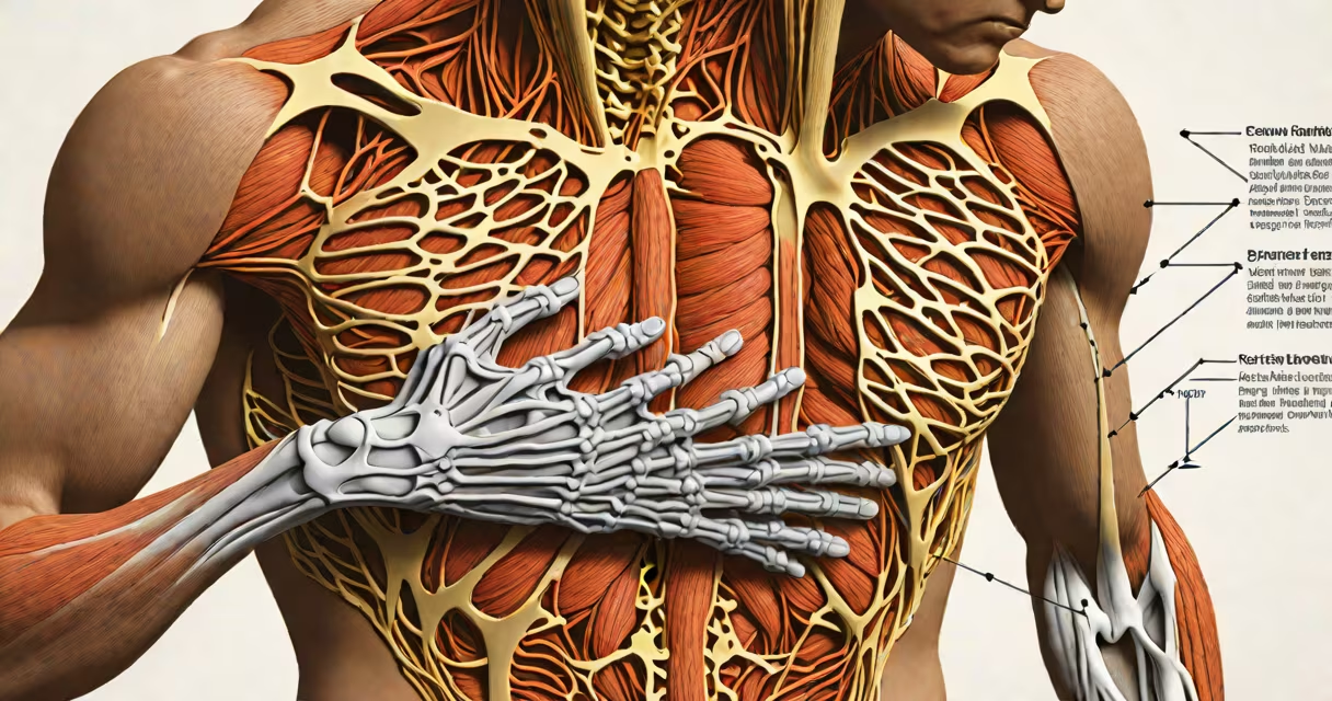
Skeletal muscle chest pain arises from strain, trauma, or inflammation in the muscles, tendons, or cartilage of the chest wall-not from the heart or lungs. It typically presents as sharp, localized pain worsened by movement, deep breathing, or pressure on the affected area.
This condition is common in people who perform repetitive upper-body motions, lift heavy objects, or experience sudden physical stress. Unlike cardiac chest pain, which may radiate to the arm or jaw, skeletal muscle chest pain stays confined to the chest wall and often improves with rest and over-the-counter anti-inflammatories.
According to the American College of Rheumatology, musculoskeletal causes account for up to 30% of non-cardiac chest pain cases seen in primary care settings. The most frequent diagnosis is costochondritis-acute inflammation of the cartilage connecting the ribs to the sternum-which affects about 1 in 200 adults annually.
The chest wall contains multiple layers of skeletal muscle, including the pectoralis major and minor, intercostal muscles between the ribs, and the rectus abdominis extending into the lower rib cage. These muscles stabilize the torso, assist in breathing, and support posture.
Injury occurs through direct impact, overuse, poor lifting mechanics, or sudden twisting motions. For example, a weightlifter performing bench presses with improper form may develop microtears in the pectoralis major, leading to localized soreness that worsens during push-ups or reaching overhead.
Pain signals originate in nociceptors-pain receptors-in the connective tissue around the muscles. These send impulses via spinal nerves to the brain, where they're interpreted as discomfort in the chest. The sensation is usually sharp, aching, or burning and may last days to weeks depending on severity.
While all skeletal muscle chest pain shares a common origin, different patterns emerge based on location and cause:
| Type | Location | Common Cause | Distinctive Feature |
|---|---|---|---|
| Costochondritis | Front of chest, near sternum | Repetitive motion, viral illness, trauma | Pain increases with deep breaths or coughing; tenderness when pressing on rib joints |
| Intercostal strain | Between ribs, along sides of chest | Sudden twist, heavy lifting, forceful sneezing | Pain worsens with side bending or rotation; may feel like a "stabbing" sensation |
| Pectoralis strain | Chest center, above the nipple line | Overexertion during exercise, sudden pull | Sharp pain during pushing movements; swelling or bruising possible |
A 2021 study published in Journal of Pain Research found that 68% of patients diagnosed with non-cardiac chest pain had identifiable musculoskeletal triggers, with costochondritis being the most prevalent.
Skeletal muscle chest pain is frequently misdiagnosed as cardiac-related due to overlapping symptoms. Both can cause tightness, pressure, or burning sensations in the chest. However, key differences exist.
Cardiac pain typically lasts longer than five minutes, may radiate to the left arm, neck, or jaw, and often occurs during exertion. Skeletal muscle pain appears suddenly after activity, is reproducible with palpation, and improves with rest and positioning changes.
One non-obvious distinction: cardiac ischemia can cause referred pain to the shoulder or jaw, while musculoskeletal pain rarely travels beyond the rib cage. A 2019 review in Annals of Internal Medicine noted that nearly half of patients presenting with chest pain initially receive a cardiac workup-even when risk factors are absent-highlighting the need for careful differential diagnosis.
I treated a 34-year-old male office worker who reported sharp chest pain after moving furniture. He described the pain as "like a knife under my left rib," worsening when he leaned forward or took deep breaths. He had no history of heart disease, smoked occasionally, and was otherwise healthy.
On exam, he had localized tenderness over the sixth rib on the left side. No swelling, redness, or warmth. He could reproduce the pain by pressing directly on the area. An EKG showed normal sinus rhythm, and blood tests for troponin were negative.
Diagnosis: left-sided intercostal muscle strain. Treatment included NSAIDs, heat therapy, and avoiding overhead reaching. Pain resolved within seven days with no recurrence after six months of follow-up.
Most cases resolve within days to two weeks with conservative management. But seek evaluation if:
These signs suggest alternative diagnoses such as pulmonary embolism, pleuritis, or cardiac ischemia. The American Heart Association recommends immediate medical attention for any new-onset chest pain with associated symptoms.
Understanding the difference between skeletal muscle chest pain and cardiac events prevents unnecessary anxiety and ensures timely treatment. When pain is localized, reproducible, and linked to movement, it is likely musculoskeletal-and manageable with targeted care.