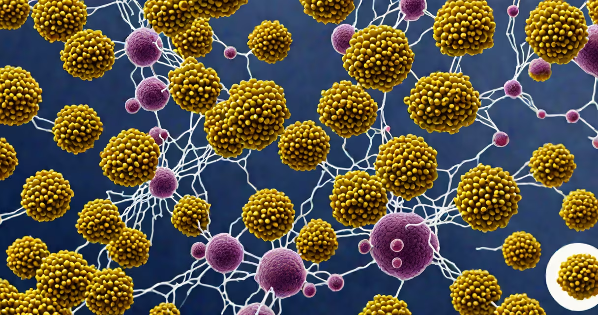
Staphylococcus aureus in lungs refers to a bacterial infection caused by the Gram-positive bacterium Staphylococcus aureus that invades lung tissue, leading to pneumonia, lung abscesses, or sepsis. It can occur in healthy individuals or severely compromise those with weakened immunity, chronic illness, or recent hospitalization.
This pathogen is responsible for both community-acquired and healthcare-associated respiratory infections. It colonizes the nose in about 30% of people, and when it breaches the respiratory tract, it triggers inflammation, pus formation, and tissue damage. The condition is particularly dangerous because some strains resist common antibiotics, including methicillin, making them harder to treat.
According to the Centers for Disease Control and Prevention (CDC), invasive S. aureus infections-including those affecting the lungs-account for over 100,000 hospitalizations annually in the United States. These cases often require intensive care and prolonged antibiotic therapy. Early detection and targeted treatment are critical to preventing death.
S. aureus enters the lungs through inhalation of contaminated droplets or by spreading from another site in the body, such as a skin wound or bloodstream infection. Once inside the airways, it adheres to epithelial cells using surface proteins like fibronectin-binding protein A. This attachment allows it to multiply rapidly and evade immune defenses.
The bacteria release toxins such as alpha-toxin and Panton-Valentine leukocidin (PVL), which destroy white blood cells and lung tissue. This leads to necrotizing pneumonia-where lung tissue dies-and can result in cavitation, abscess formation, or empyema (pus in the pleural space).
A 2021 study published in Clinical Infectious Diseases found that patients with S. aureus pneumonia had a 30-day mortality rate of 24%, higher than other bacterial pneumonias. Risk factors include mechanical ventilation, prior antibiotic use, and underlying conditions like COPD or diabetes.
S. aureus in lungs manifests in several clinical patterns depending on patient health and exposure:
| Type | Description | Common Setting | Key Features |
|---|---|---|---|
| Community-acquired pneumonia (CAP) | Occurs in otherwise healthy people, often after viral upper respiratory infection | Outpatient clinics, emergency rooms | Fever, productive cough, rapid breathing; may progress to septic shock |
| Hospital-acquired pneumonia (HAP) | Develops 48 hours or more after hospital admission | ICU, post-surgical wards | Often linked to ventilators; high mortality due to multidrug-resistant strains |
| Methicillin-resistant S. aureus (MRSA) pneumonia | Resistant to beta-lactam antibiotics; requires vancomycin or linezolid | Long-term care facilities, trauma centers | More likely to cause necrotizing changes and lung abscesses |
| Bacteremic pneumonia | Spread from bloodstream infection; often presents with fever and sepsis | Patients with IV drug use history or prosthetic devices | May show multiple lung nodules on CT scan |
One non-obvious fact: S. aureus can survive for days on dry surfaces in hospitals. A 2019 study in Infection Control & Hospital Epidemiology showed the bacterium remained viable on bed rails and stethoscopes for up to 7 days, contributing to nosocomial transmission.
Diagnosing S. aureus in lungs begins with imaging. Chest X-rays often show infiltrates, while CT scans reveal cavitary lesions or pleural effusions. Definitive diagnosis requires culture of sputum, bronchoalveolar lavage fluid, or blood. However, contamination during sample collection can lead to false positives.
Antibiotic selection hinges on susceptibility testing. For methicillin-sensitive strains, nafcillin or oxacillin are first-line. For MRSA, vancomycin or daptomycin is used. Treatment duration ranges from 2 to 6 weeks, depending on severity and response.
I once observed a case in a medical rotation where a 68-year-old man with diabetes developed sudden hypoxia after a flu-like illness. His chest CT showed bilateral infiltrates with a right-sided cavity. Sputum culture grew MRSA. He required intubation and intravenous vancomycin. After 21 days of therapy, he recovered-but not before developing a lung abscess requiring drainage.
S. aureus in lungs is frequently mistaken for viral pneumonia or fungal infections. Viral pneumonia typically presents with a gradual onset, lymphocytosis on blood tests, and normal chest X-ray early on. Fungal pneumonia (e.g., from Aspergillus) tends to affect immunocompromised hosts and shows a "halo sign" on CT.
Another key distinction: S. aureus pneumonia often causes rapid clinical deterioration within 24-48 hours. Patients may go from stable to requiring ICU admission quickly. This contrasts with typical bacterial pneumonia, which progresses over days.
Some clinicians confuse S. aureus with Streptococcus pneumoniae. While both cause lobar pneumonia, S. aureus is more likely to produce pleural effusion, empyema, and necrosis. It also responds poorly to penicillin and cephalosporins unless the strain is sensitive.
Preventing S. aureus in lungs involves decolonization protocols in high-risk settings. The CDC recommends screening and nasal mupirocin application for patients undergoing surgery or admitted to ICUs. Hand hygiene and environmental cleaning reduce transmission.
Despite advances, outcomes remain poor for severe cases. Mortality exceeds 20% in hospitalized patients with MRSA pneumonia. Early recognition and aggressive management improve survival. Delayed treatment increases risk of complications like respiratory failure or death.