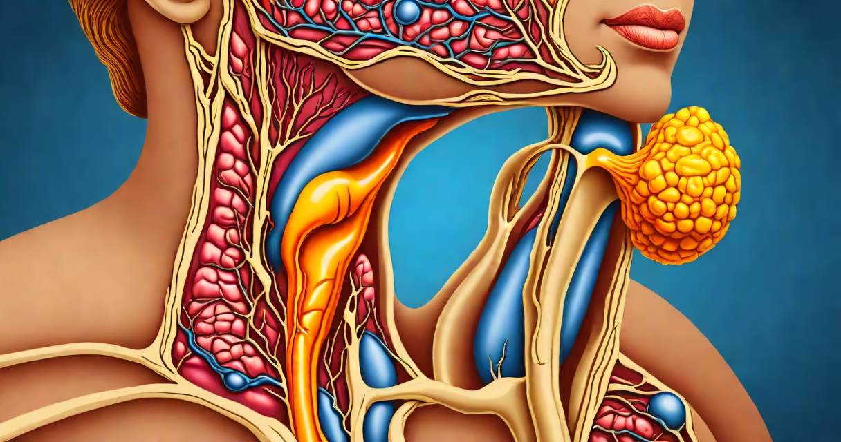
A thyroid tumor is an abnormal mass of tissue that forms within the thyroid gland, a butterfly-shaped organ located at the base of the neck that regulates metabolism through hormone production. These growths can be noncancerous (benign) or cancerous (malignant), with most being detected incidentally during imaging for unrelated issues.
Thyroid tumors develop when cells in the thyroid tissue grow uncontrollably. The majority-about 80%-are benign nodules, often discovered during routine physical exams or imaging scans such as ultrasound. Malignant tumors, while less common, account for approximately 1% of all cancers diagnosed annually in the U.S., according to the American Cancer Society.
These tumors originate from different cell types in the thyroid. The most frequent subtype is papillary thyroid carcinoma, which arises from follicular cells and accounts for about 80% of all thyroid cancers. Other types include follicular, medullary, and anaplastic thyroid cancers, each with distinct genetic profiles and clinical behaviors.
Thyroid tumors begin when mutations accumulate in thyroid cells. Common triggers include radiation exposure, especially in childhood, and inherited genetic conditions such as familial medullary thyroid cancer (FMTC) or multiple endocrine neoplasia type 2 (MEN2).
Detection typically occurs through physical exam, ultrasound, or blood tests. A doctor may feel a lump during a neck palpation. Ultrasound uses sound waves to visualize internal structures and can distinguish solid from fluid-filled nodules. Nodules larger than 1 cm are usually biopsied using fine-needle aspiration (FNA), where a thin needle extracts cells for microscopic analysis.
For example, a 45-year-old woman with no symptoms underwent a routine CT scan for abdominal pain. The scan revealed a 1.3 cm solid nodule in her right lobe. An FNA biopsy confirmed it was a papillary thyroid carcinoma. She later underwent surgical removal of the affected lobe and remains under follow-up care.
Thyroid tumors are classified by histology-the appearance of cells under a microscope-and their biological behavior. The following table outlines major types:
| Type | Prevalence | Prognosis | Key Features |
|---|---|---|---|
| Papillary | ~80% | Excellent (95% 10-year survival) | Slow-growing, often spreads to lymph nodes, responds well to treatment |
| Follicular | ~10-15% | Good (90% 10-year survival) | Less likely to spread to lymph nodes; more prone to distant metastasis |
| Medullary | ~3-5% | Moderate (80% 10-year survival) | Arises from calcitonin-producing C-cells; associated with MEN2 syndrome |
| Anaplastic | <1% | Poor (median survival <6 months) | Rapidly growing, aggressive; rarely curable |
One non-obvious distinction: not all thyroid tumors cause symptoms. Many remain asymptomatic until they reach a size that compresses nearby structures. For instance, a tumor pressing on the trachea may cause hoarseness or breathing difficulty. A 2021 study published in Thyroid found that 60% of patients with papillary thyroid cancer reported no symptoms at diagnosis.
People often confuse thyroid tumors with thyroid nodules. While all tumors are nodules, not all nodules are tumors. A nodule is simply a lump in the thyroid, regardless of whether it's solid, fluid-filled, or benign. A tumor implies abnormal cellular proliferation, but not necessarily malignancy.
Another misconception is that a thyroid tumor always requires surgery. This is false. Small, benign-appearing nodules may be monitored with periodic ultrasounds and blood tests. The American Thyroid Association recommends observation for nodules under 1 cm with low-risk ultrasound features.
I once observed a patient with a 0.8 cm nodule showing microcalcifications on ultrasound-a red flag. Despite the concerning appearance, the FNA result was benign. The patient was reassured and followed with annual imaging. This case underscores that imaging findings alone don't confirm cancer.
Medical evaluation begins when a nodule is detected or symptoms arise. Symptoms prompting concern include persistent hoarseness, difficulty swallowing, neck swelling, or a rapidly growing lump. Blood tests measuring TSH, free T4, and thyroglobulin levels help assess thyroid function and tumor activity.
Biopsy results determine the next step. If benign, monitoring is standard. If suspicious or malignant, treatment options include thyroidectomy (removal of part or all of the thyroid), radioactive iodine therapy, and thyroid hormone suppression therapy to lower TSH and reduce tumor recurrence risk.
For example, a 52-year-old man with a 2.1 cm nodule and enlarged cervical lymph nodes underwent total thyroidectomy. Pathology confirmed stage II papillary thyroid cancer. He received radioactive iodine ablation and now takes levothyroxine to suppress TSH below 0.1 mIU/L, a strategy shown to reduce recurrence rates by 40% in long-term studies.
Not all thyroid tumors progress. Some remain stable for years. However, early detection improves outcomes. The National Cancer Institute reports that localized thyroid cancer has a 98% five-year survival rate, compared to 55% for distant-stage disease.
Understanding what is a thyroid tumor involves recognizing that it's not a single condition but a group of disorders with varying risks and treatments. Early detection through screening and informed decision-making based on pathology and imaging are key to managing them effectively.