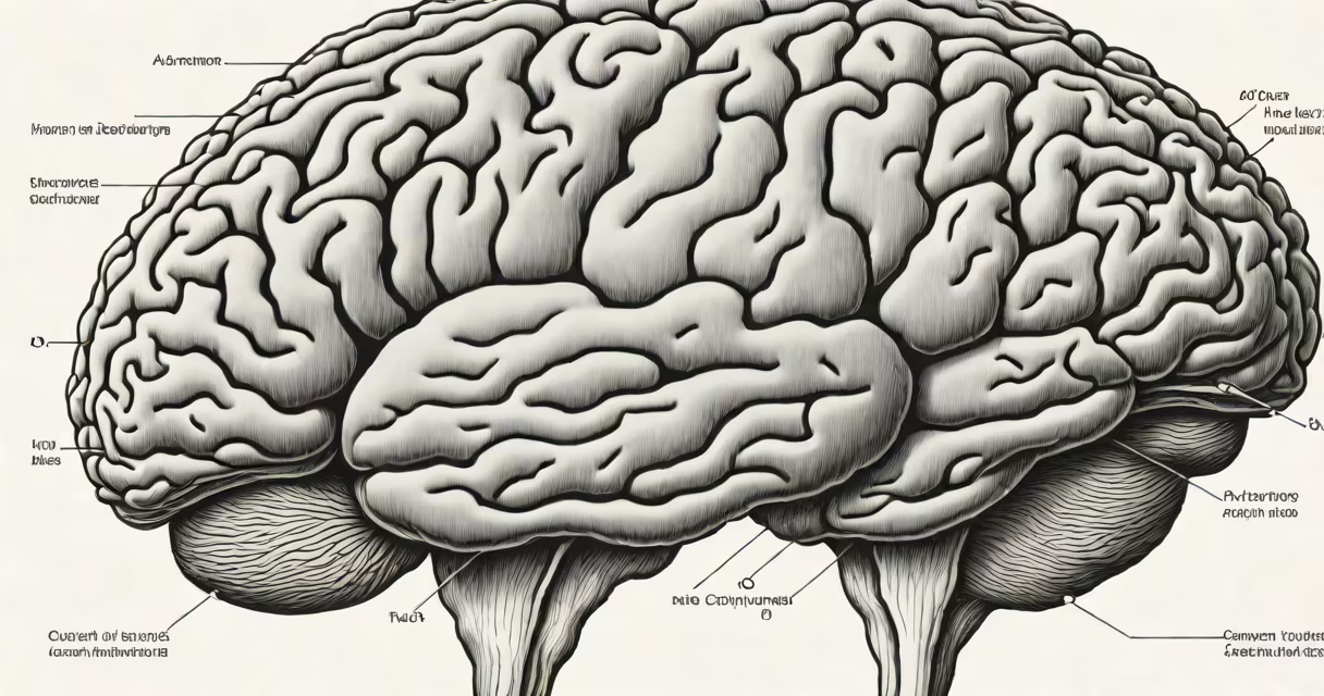
Brain swelling in adults, medically known as cerebral edema, occurs when excess fluid accumulates in brain tissue, increasing intracranial pressure and potentially causing life-threatening complications. This condition arises from trauma, stroke, infections, tumors, or metabolic disturbances.
Cerebral edema develops when the blood-brain barrier becomes compromised or when cellular energy failure disrupts ion balance. The resulting fluid shift into brain cells or extracellular spaces increases volume within the rigid skull. Even small increases in pressure can compress important structures, impair blood flow, and lead to herniation-a fatal condition where brain tissue shifts through natural openings in the skull.
According to the National Institute of Neurological Disorders and Stroke (NINDS), cerebral edema affects approximately 10% of patients with severe traumatic brain injury and up to 30% of those with ischemic stroke. Early recognition and intervention are essential to prevent irreversible damage.
Swelling begins when brain cells fail to regulate water and electrolytes properly. Normally, astrocytes-supportive glial cells-maintain osmotic balance. When oxygen supply drops during hypoxia or ischemia, ATP production halts. Sodium-potassium pumps stop working, sodium accumulates inside cells, and water follows passively via osmosis.
This process, called cytotoxic edema, causes neurons and glial cells to swell. It typically appears within minutes of injury. Over time, vascular damage allows plasma proteins and fluid to leak into the extracellular space-called vasogenic edema. This form often peaks 24-72 hours after injury and contributes to mass effect and elevated pressure.
A third type, interstitial edema, occurs in obstructive hydrocephalus when cerebrospinal fluid (CSF) builds up and pushes fluid into brain tissue. This is less common in adults but still relevant in cases of brain tumors or congenital abnormalities.
Several conditions trigger brain swelling in adults. Traumatic brain injury (TBI) accounts for nearly half of all acute cerebral edema cases. A fall, motor vehicle accident, or assault can cause diffuse axonal injury or contusions, leading to immediate swelling. For example, a 42-year-old man involved in a motorcycle crash sustained a subdural hematoma and developed progressive swelling over 12 hours, requiring emergency craniotomy.
Ischemic stroke causes cytotoxic edema within 1-2 hours of blocked blood flow. The penumbra-the area around the core infarct-can swell rapidly. Studies show that up to 60% of large hemispheric strokes result in significant edema, especially in middle-aged men with hypertension and diabetes.
Infections such as bacterial meningitis or viral encephalitis also induce swelling. Herpes simplex encephalitis, for instance, targets temporal lobes and causes focal swelling. A 2021 study published in The Lancet Neurology reported that 43% of patients with herpes encephalitis developed cerebral edema requiring ICU management.
Tumors-particularly high-grade gliomas-trigger vasogenic edema. As tumor cells invade surrounding tissue, they disrupt capillary integrity. Steroid therapy (e.g., dexamethasone) reduces edema in about 70% of cases, but swelling may persist or worsen despite treatment.
| Condition | Primary Edema Type | Onset Time | Key Risk Factors |
|---|---|---|---|
| Traumatic brain injury | Cytotoxic & vasogenic | Minutes to hours | High-impact trauma, alcohol use, age >65 |
| Ischemic stroke | Cytotoxic | 1-2 hours | Hypertension, atrial fibrillation, prior stroke |
| Bacterial meningitis | Vasogenic | Hours to days | Immune suppression, recent surgery, CSF leak |
| Glioblastoma | Vasogenic | Days to weeks | Age 50+, history of cancer, genetic mutations |
Brain swelling is often mistaken for general headache or fatigue. However, symptoms like worsening headache, confusion, nausea, vomiting, seizures, or altered consciousness signal neurological deterioration. A sudden decline in Glasgow Coma Scale (GCS) score-especially below 8-indicates urgent need for imaging.
CT scans and MRI are standard tools. On CT, swelling appears as loss of gray-white matter differentiation, midline shift, and compressed ventricles. MRI shows hyperintensity on T2/FLAIR sequences. A key diagnostic clue is the presence of diffusion restriction on DWI sequences, indicating cytotoxic edema.
One common error is assuming swelling always correlates with visible lesions. In some cases, such as post-hypoxic encephalopathy after cardiac arrest, swelling occurs without structural damage. A 2020 study in JAMA Neurology found that 28% of patients with normal initial MRI scans later developed significant edema during hospitalization.
A 61-year-old woman presented with left-sided weakness and aphasia. An initial CT scan showed no hemorrhage but revealed early signs of right middle cerebral artery territory infarction. Within six hours, her GCS dropped from 15 to 10. Repeat imaging confirmed massive swelling in the right frontal and parietal lobes, with midline shift of 1.2 cm.
She was treated with IV mannitol (0.5 g/kg) and corticosteroids. Despite this, her intracranial pressure rose above 25 mmHg. A decompressive craniectomy was performed, removing part of the skull to relieve pressure. Her recovery took eight weeks, but she regained speech and partial mobility. This case illustrates how rapid progression demands immediate neurosurgical intervention.
Brain swelling is not the same as pseudotumor cerebri (idiopathic intracranial hypertension), which primarily affects young obese women and causes increased CSF pressure without actual tissue swelling. It lacks focal deficits and responds to lumbar puncture and acetazolamide.
Also distinct is cerebral venous sinus thrombosis (CVST), which causes swelling due to impaired drainage rather than direct tissue injury. CVST presents with headaches, seizures, and papilledema. MRI with MRV confirms the diagnosis. Unlike edema from stroke, CVST swelling resolves with anticoagulation.
Another frequent mix-up is confusing brain swelling with brain abscess. While both cause mass effect, abscesses contain pus and show ring-enhancing lesions on contrast MRI. They require antibiotics and surgical drainage, not just osmotic agents.
I observed a patient with a ruptured aneurysm who developed rapid cerebral edema post-surgery. Despite normal blood pressure and oxygenation, his pupils became asymmetric. A follow-up CT scan showed diffuse swelling and midline shift. We administered hypertonic saline and initiated hyperventilation. After 48 hours, swelling stabilized. This case underscored that even after successful surgery, secondary swelling remains a major threat.
Prevention focuses on managing risk factors: controlling blood pressure, treating infections early, and preventing head trauma. In trauma settings, rapid stabilization and early imaging reduce delayed edema formation.
Treatment options depend on severity. Mild cases may respond to osmotherapy (mannitol or hypertonic saline), steroids (for vasogenic edema), or hypothermia. Severe cases require surgical decompression. Craniectomy reduces mortality by 20% in patients with malignant edema, according to a 2019 Cochrane review.
Monitoring intracranial pressure via an intraventricular catheter is standard in intensive care. Target pressure should remain below 20 mmHg. Continuous EEG monitoring detects seizures that worsen edema and guide anticonvulsant use.
Understanding what causes brain swelling in adults enables faster diagnosis and targeted therapy. Early action prevents irreversible damage and improves outcomes. Recognizing subtle signs-like persistent vomiting or worsening confusion-can save lives.