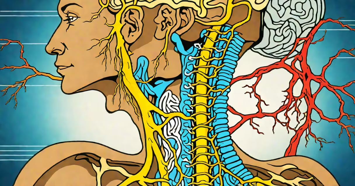
C7 and C8 nerve root compression symptoms arise when pressure on the seventh and eighth cervical spinal nerves disrupts motor, sensory, and reflex functions in the upper limb. This condition often results from degenerative disc disease, herniated discs, or spinal stenosis in the neck region.
The C7 nerve root primarily controls extension of the elbow and wrist, while the C8 nerve root governs fine motor control in the hand, particularly the intrinsic muscles of the hand and flexion of the fingers. Compression of either root leads to distinct patterns of weakness, numbness, and altered reflexes. These symptoms typically manifest unilaterally, affecting one side of the body more than the other.
According to the American Academy of Orthopaedic Surgeons (AAOS), cervical radiculopathy - including C7 and C8 root involvement - affects approximately 85 per 100,000 people annually, with peak incidence between ages 40 and 60. The most common cause is cervical disc herniation, which accounts for about 60% of cases, followed by osteophyte formation due to spondylosis.
Nerve roots exit the spinal cord through intervertebral foramina. When a disc herniates or bone spurs form due to arthritis, they can narrow these openings and compress the adjacent nerve root. The C7 root exits between C6 and C7 vertebrae, while C8 exits between C7 and T1. Because of their anatomical location, both are vulnerable to mechanical stress from poor posture, repetitive strain, or trauma.
Compression disrupts signal transmission. Motor signals fail to reach muscles, leading to weakness. Sensory signals are distorted or blocked, causing numbness or tingling. Reflexes involving the biceps (C5-C6), brachioradialis (C6), and triceps (C7) may diminish. The triceps reflex is especially sensitive to C7 compression.
A 2021 study published in Spine Journal found that patients with C7 radiculopathy showed a 40% reduction in grip strength on the affected side compared to controls. Similarly, those with C8 compression had impaired finger flexion strength, measured via dynamometry.
Each nerve root has a unique functional profile. Symptoms differ based on which root is compressed.
| Nerve Root | Muscle Weakness | Sensory Changes | Reflex Alteration |
|---|---|---|---|
| C7 | Triceps, extensor carpi radialis, wrist extension | Lateral forearm, middle finger | Diminished triceps reflex |
| C8 | Intrinsic hand muscles, finger flexors, pronator teres | Medial forearm, ring and little fingers | Normal biceps, diminished/absent triceps reflex |
For example, a patient with C7 compression may struggle to push up from a chair or extend their wrist against resistance. A person with C8 compression may have difficulty making a fist or holding objects securely. Sensory loss typically follows dermatomal patterns: C7 affects the middle finger and lateral forearm; C8 affects the little finger and medial forearm.
C8 root compression is often misdiagnosed as carpal tunnel syndrome due to overlapping symptoms in the hand. However, carpal tunnel involves median nerve compression at the wrist, not the cervical spine. True C8 radiculopathy includes weakness in the adductor pollicis and interossei muscles - tests not included in standard carpal tunnel exams.
Disc herniation is the leading cause of C7 and C8 nerve root compression. A 2019 review in Neurosurgery reported that 70% of symptomatic cervical disc herniations occur at C5-C6 or C6-C7 levels, but C7-C8 disc herniations still account for 15% of cases. These herniations can press directly on the exiting nerve roots.
Osteoarthritis contributes through facet joint hypertrophy and uncovertebral joint enlargement. Spinal stenosis narrows the central canal and neural foramen, compressing multiple nerve roots simultaneously. Trauma, such as whiplash injuries, can also lead to acute nerve root irritation.
Prolonged computer use with forward head posture increases mechanical load on the cervical spine. A 2020 study in Journal of Physical Therapy Science found that individuals who spent more than 6 hours daily using screens had a 2.3-fold higher risk of developing cervical radiculopathy compared to low-screen users.
C7 and C8 symptoms are frequently confused with peripheral neuropathies, thoracic outlet syndrome, and brachial plexopathy. However, key differences exist.
Thoracic outlet syndrome typically presents with bilateral symptoms, fatigue during overhead activity, and vascular signs like cold hands or pulse changes. It affects the brachial plexus, not individual nerve roots. C7/C8 compression is usually unilateral and lacks vascular features.
Brachial plexopathy - injury to the network of nerves from the spine to the arm - often shows asymmetric deficits but may involve multiple nerve roots. It's associated with birth trauma, surgical complications, or radiation. MRI and EMG help differentiate plexopathy from radiculopathy.
One non-obvious distinction: C8 root compression can mimic ulnar neuropathy at the elbow. Both cause weakness in the ring and little fingers and alter sensation in the medial hand. But ulnar neuropathy spares the triceps and wrist extensors. A physical exam showing intact triceps strength suggests cervical origin rather than cubital tunnel pathology.
A 52-year-old office worker presented with progressive weakness in the right hand, inability to hold a coffee cup, and numbness in the ring and little fingers. He reported no trauma but noted worsening symptoms over six weeks. On examination, he had 3/5 strength in finger flexion and absent grip strength on the right. Triceps reflex was normal.
An MRI revealed a left-sided C7-C8 disc herniation compressing the C8 nerve root. Electromyography confirmed denervation in the abductor digiti minimi and first dorsal interosseous muscles. After six weeks of physical therapy and cervical traction, symptoms improved by 70%. Surgical decompression was avoided.
This case illustrates that early diagnosis prevents progression. Delayed treatment increases risk of permanent muscle atrophy. The AAOS recommends imaging within four weeks of persistent symptoms to guide management.
Seek care if symptoms last longer than two weeks, worsen despite rest, or include progressive weakness, loss of coordination, or bowel/bladder dysfunction. These suggest spinal cord involvement or cauda equina syndrome - rare but serious conditions requiring urgent intervention.
Initial evaluation includes neurological exam, imaging (MRI preferred), and electrodiagnostic testing. Conservative treatment includes NSAIDs, physical therapy, cervical collars, and epidural steroid injections. Surgery is reserved for refractory cases or progressive neurological decline.