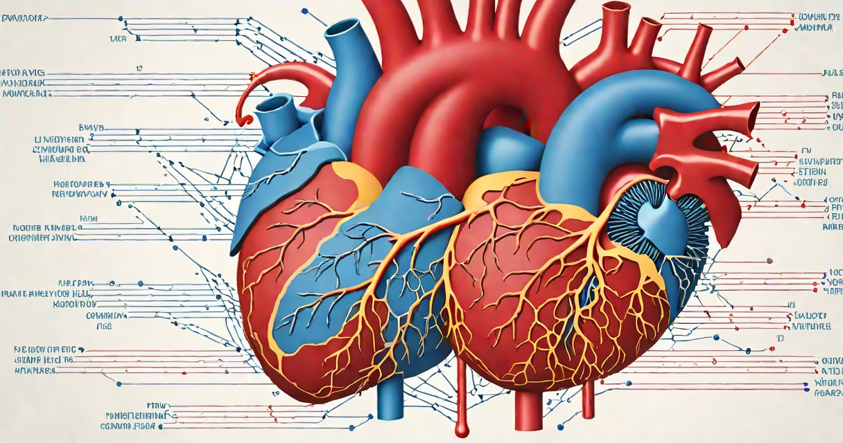
The flow of the heart is the continuous movement of blood through the heart's four chambers and associated vessels, powered by coordinated contractions of cardiac muscle. This process delivers oxygen-rich blood to tissues and returns deoxygenated blood to the lungs for re-oxygenation.
Each heartbeat begins with electrical signals originating in the sinoatrial node, triggering atrial contraction. Blood flows from the atria into the ventricles during diastole. Then, ventricular systole forces blood out through the pulmonary artery and aorta. The entire cycle repeats about 60-100 times per minute in adults at rest.
According to the American Heart Association, the average adult heart pumps approximately 7,500 liters of blood daily-enough to fill a small swimming pool. This volume underscores the efficiency required in maintaining consistent flow under varying physiological demands.
Blood enters the right atrium via the superior and inferior vena cava, carrying carbon dioxide from systemic circulation. It passes through the tricuspid valve into the right ventricle. During ventricular contraction, the pulmonary valve opens, sending blood to the lungs.
In the lungs, gas exchange occurs: carbon dioxide exits, oxygen binds to hemoglobin. Oxygenated blood returns to the left atrium via the pulmonary veins. It flows through the mitral valve into the left ventricle. Contraction of the left ventricle ejects blood into the aorta, distributing oxygen to the brain, muscles, and organs.
Valves prevent backflow. The tricuspid and mitral valves close during ventricular contraction to stop regurgitation. The pulmonary and aortic valves snap shut after ejection, preventing blood from flowing backward into the ventricles.
The flow of the heart involves both laminar and turbulent patterns. Laminar flow occurs smoothly along vessel walls, minimizing resistance. Turbulent flow develops when blood velocity increases sharply-such as at valve openings or narrowed arteries-and can produce audible sounds detectable with a stethoscope.
| Type | Description | Example |
|---|---|---|
| Laminar | Smooth, layered flow; low resistance | Normal blood flow in healthy arteries |
| Turbulent | Chaotic, eddying motion; higher resistance | Flow near stenosed coronary arteries |
| Reversed | Backward flow due to valve failure | Regurgitation in mitral insufficiency |
When turbulence exceeds normal thresholds, it generates murmurs. A physician may hear a systolic murmur at the left lower sternal border indicating pulmonic stenosis. These sounds are diagnostic clues to underlying structural issues.
Non-obvious fact: Flow dynamics are not uniform across the heart's chambers. The left ventricle generates pressures up to 120 mmHg during systole-over four times higher than the right ventricle's 25 mmHg. This difference reflects the greater resistance of systemic circulation compared to pulmonary circulation.
Consistent flow prevents tissue hypoxia. Without adequate perfusion, cells switch to anaerobic metabolism, producing lactic acid and leading to organ dysfunction. In the brain, even five minutes without sufficient blood flow causes irreversible damage.
Cardiac output-the volume of blood pumped per minute-is calculated as stroke volume × heart rate. A typical adult has a resting cardiac output of 5-6 liters per minute. During exercise, this can increase to 20-25 liters per minute, reflecting changing regulation of flow.
Abnormal flow patterns indicate disease. For example, a reduced ejection fraction (less than 40%) signals heart failure. Echo Doppler imaging measures flow velocity and direction, allowing clinicians to quantify valvular regurgitation or assess shunts in congenital defects.
Many assume the heart "pumps" blood like a mechanical pump. But the heart is a biological engine relying on elastic recoil and pressure gradients. Its walls stretch during filling and contract during ejection-not unlike a water balloon squeezed by hand.
Another misconception: blood flows in a single loop. In reality, two circuits exist-the pulmonary and systemic. Deoxygenated blood travels from the body to the lungs, then returns oxygenated to the heart and is distributed to the body. This dual circuit design separates high-pressure systemic flow from low-pressure pulmonary flow.
During my time working in a cardiology clinic, I observed a patient with severe aortic stenosis. His echocardiogram showed peak flow velocity of 4.8 m/s across the aortic valve-well above the normal threshold of 1.3 m/s. This indicated significant obstruction, requiring surgical intervention. The abnormal flow pattern was visible in real-time color Doppler imaging.
Flow of the heart differs from pulse, which is the palpable expansion of arteries caused by each heartbeat. Pulse rate usually equals heart rate, but discrepancies occur in arrhythmias. Atrial fibrillation, for example, may cause irregular pulses despite a steady heart rate.
Flow also differs from blood pressure. Pressure is force per unit area exerted on vessel walls. Flow is volume per unit time. High pressure doesn't always mean high flow-narrowed vessels restrict flow even if pressure remains elevated.
Understanding the flow of the heart reveals how biology maintains life through precise, automated mechanics. It is not a single event but a continuous, adaptive process essential to survival.