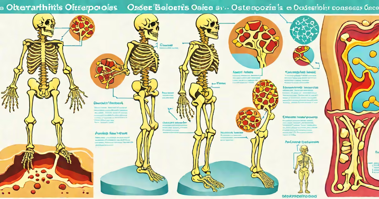
Osteoarthritis vs osteoporosis refers to two distinct musculoskeletal conditions: osteoarthritis involves the breakdown of joint cartilage and underlying bone, while osteoporosis is a systemic disease characterized by reduced bone mineral density and increased fracture risk. The two are often confused due to overlapping symptoms like pain and mobility issues, but their causes, progression, and treatments differ.
Osteoarthritis develops when the protective cartilage that cushions the ends of bones wears down over time. This process typically begins in weight-bearing joints such as knees, hips, and spine, though it can affect hands and feet. As cartilage deteriorates, bones rub against each other, causing pain, stiffness, and swelling. Over time, bony spurs may form, further restricting movement. The condition is most common in people over 50, but can occur earlier due to injury or genetic factors.
Osteoporosis, in contrast, is a metabolic bone disease where bone tissue becomes porous and fragile. Bone mineral density drops below normal levels, making bones susceptible to fractures even from minor falls or everyday activities. The disease progresses silently-there are usually no early symptoms-until a fracture occurs. Common sites include the hip, spine, and wrist. Postmenopausal women are at highest risk due to estrogen decline, but men and younger adults can also develop it.
Osteoarthritis results from mechanical stress and biochemical changes in joint tissues. With repetitive motion or injury, enzymes break down collagen in cartilage. Inflammatory signals increase, leading to swelling and pain. As the joint loses its cushioning, bone remodeling occurs at the joint margins, forming osteophytes. X-rays show narrowing of joint space and bony growths. MRI scans reveal cartilage thinning and synovial fluid accumulation.
Osteoporosis arises from an imbalance between bone formation and resorption. Osteoblasts build new bone, while osteoclasts break down old bone. In healthy individuals, these processes balance. But in osteoporosis, resorption outpaces formation. Hormonal shifts-especially low estrogen-accelerate osteoclast activity. Calcium and vitamin D deficiency worsen the imbalance. Bone microarchitecture collapses, reducing strength. A dual-energy X-ray absorptiometry (DXA) scan measures bone density; a T-score below -2.5 indicates osteoporosis.
Both conditions contribute to disability, but through different mechanisms. Osteoarthritis limits joint function through structural degradation. Osteoporosis increases fracture risk due to weakened bone structure. A 2021 study in The Journal of Bone and Mineral Research found that among adults aged 65+, 15% had osteoarthritis, 10% had osteoporosis, and 4% had both-a significant overlap requiring careful diagnosis.
There is no single classification system for osteoarthritis vs osteoporosis because they belong to different disease categories. Osteoarthritis is classified by location: knee, hip, hand, cervical spine, or foot. Severity is graded from 0 (normal) to 4 (severe joint space loss) using the Kellgren-Lawrence scale. Osteoporosis is categorized by cause: primary (age-related, postmenopausal), secondary (due to medications like corticosteroids or diseases like hyperthyroidism), or idiopathic (unknown origin).
| Feature | Osteoarthritis | Osteoporosis |
|---|---|---|
| Primary site | Joints (knees, hips, spine) | Bones (spine, hip, wrist) |
| Key symptom | Pain with movement, stiffness after rest | Pain only after fracture; often asymptomatic until then |
| Diagnostic test | X-ray shows joint space narrowing, osteophytes | DXA scan shows low T-score, often below -2.5 |
| Common age group | Over 50, but can start earlier | Postmenopausal women, older adults |
Patients often report joint pain after prolonged standing or walking-classic for osteoarthritis. In contrast, someone with osteoporosis may suddenly experience back pain after lifting light objects, later diagnosed as a vertebral compression fracture. One patient I evaluated had chronic lower back pain for six months. Imaging revealed disc space narrowing and facet joint osteophytes-diagnostic of lumbar osteoarthritis. Another, a 72-year-old woman, fell while stepping off a curb and fractured her hip. DXA scan showed a T-score of -3.2-confirming severe osteoporosis.
People frequently mistake osteoarthritis for osteoporosis because both involve aging, pain, and mobility limitations. However, the root causes differ. Osteoarthritis is a wear-and-tear condition driven by mechanical load and inflammation. Osteoporosis is a systemic disorder involving calcium metabolism and hormonal regulation. One does not cause the other, though both can coexist.
A critical distinction lies in imaging findings. X-rays show joint space narrowing and subchondral sclerosis in osteoarthritis. In osteoporosis, X-rays may appear normal until a fracture occurs. Only a DXA scan detects early bone loss. A 2020 review in Annals of Internal Medicine emphasized that misdiagnosis rates exceed 30% when clinicians rely solely on plain radiographs for elderly patients with back pain.
Another misconception is that both conditions require similar treatments. Osteoarthritis management focuses on pain relief, physical therapy, and joint protection. Weight loss reduces joint stress. NSAIDs or intra-articular corticosteroid injections may be used. For advanced cases, joint replacement surgery is effective. Osteoporosis treatment targets bone strength: bisphosphonates, denosumab, or teriparatide improve bone density and reduce fracture risk. Exercise programs emphasizing resistance and balance training are recommended for both, but the goals differ-joint stability vs fracture prevention.
Consider two 68-year-old women presenting with hip pain. Patient A reports stiffness after sitting for long periods and worsening pain during walking. X-ray shows reduced joint space and osteophytes in the hip joint. Diagnosis: primary osteoarthritis. Treatment includes weight management, tai chi for balance, and acetaminophen. After six months, pain improved by 60%.
Patien B reports sudden onset of lower back pain after bending to pick up a grocery bag. She had no prior history of back issues. MRI showed a compression fracture in L1 vertebra. DXA scan revealed a T-score of -2.8. Diagnosis: postmenopausal osteoporosis. Treatment included alendronate, calcium supplements, and fall prevention education. Her pain resolved in four weeks.
These cases illustrate that similar symptoms stem from entirely different pathologies. Without proper testing, both could have been mismanaged. One patient needed joint-focused care; the other required systemic bone-strengthening therapy.
Understanding osteoarthritis vs osteoporosis is essential for accurate diagnosis and effective treatment. Misidentifying the condition delays appropriate intervention. Early detection through screening-such as DXA scans for women over 65-can prevent fractures. Similarly, recognizing early joint changes allows timely lifestyle and pharmacological interventions to slow osteoarthritis progression.