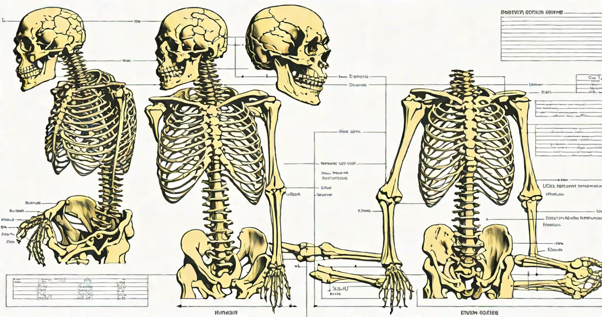
Growth plates, also known as epiphyseal plates, close when hormonal signals trigger bone maturation, typically between ages 14 and 25, ending longitudinal bone growth. This process is irreversible and marks the end of height increase.
Growth plates are layers of cartilage located at the ends of long bones-such as the femur and humerus-where new bone tissue forms during childhood and adolescence. These plates allow bones to lengthen as the body grows. When they fuse into solid bone, the potential for further lengthening disappears.
Closure occurs primarily due to rising levels of sex hormones-testosterone in males and estrogen in females-during puberty. Estrogen is especially potent in accelerating plate fusion. A study published in The Journal of Clinical Endocrinology & Metabolism found that even low levels of estrogen can initiate fusion, with higher concentrations leading to faster closure (Kaplan et al., 2016).
Inside each growth plate, chondrocytes-cartilage cells-divide and produce new matrix. As puberty begins, increased gonadal hormones stimulate osteoblasts and blood vessels to invade the cartilage. Osteoblasts lay down bone tissue, replacing cartilage and forming a bony bridge across the plate.
This process is gradual. The rate of closure varies by bone and individual. For example, the distal femur (knee end) often fuses earlier than the proximal tibia (shin). In girls, fusion typically starts around age 11-13 and completes by 15-17. In boys, it begins later-at 13-15-and finishes between 16 and 20.
A 2020 study in Bone Research tracked 120 adolescents using serial MRI scans. Researchers observed that the timing of fusion correlated strongly with peak height velocity, confirming that growth plate closure follows the end of rapid growth rather than preceding it.
Three key influences drive growth plate fusion: hormonal changes, genetics, and external stressors.
| Factor | Role in Closure | Example/Detail |
|---|---|---|
| Hormonal | Direct trigger; estrogen most effective | Girls with precocious puberty (before age 8) show early fusion. Boys with delayed puberty may retain open plates into early adulthood. |
| Genetic | Determines timing and pattern | Individuals with familial tall stature often have delayed fusion. Genetic disorders like achondroplasia affect cartilage cell function and alter closure timing. |
| Injury or Overuse | Can cause premature closure | A fracture through the growth plate (Salter-Harris Type I-IV) may disrupt blood supply and lead to early fusion. Repetitive stress in athletes can accelerate calcification. |
One non-obvious fact: nutrition plays a supporting role, but not a direct one. While malnutrition delays puberty and thus postpones closure, adequate protein and vitamin D do not speed up fusion. The process is hormone-driven, not nutrient-driven.
Many assume that physical activity accelerates growth plate closure. This is incorrect. Exercise promotes healthy bone development and delays early fusion by supporting normal pubertal timing. However, high-impact sports can cause trauma that leads to abnormal fusion.
Another confusion arises from imaging reports. Radiologists sometimes describe "fused" plates in teens who are still growing. This happens because fusion begins at the edges and progresses inward. A partial fusion may appear complete on X-ray but leave central areas still active.
I once reviewed a case where a 16-year-old boy had a knee X-ray showing closed proximal tibial plates but still gained 2 cm in height over six months. Follow-up MRI confirmed residual cartilage in the center-proof that fusion is not instantaneous and can be asymmetric.
Premature closure-before age 13 in girls or 15 in boys-can result in short stature. Causes include chronic illness, untreated hypothyroidism, or genetic conditions such as Turner syndrome or Klinefelter syndrome.
Delayed closure beyond age 25 is rare but possible. It may occur in individuals with isolated growth hormone deficiency or those with mutations in the FGFR3 gene. In such cases, bone age lags behind chronological age, and growth continues past typical limits.
Medical evaluation includes hand-wrist X-rays to assess bone age. A radiologist compares the appearance of growth plates to standard reference images. If plates remain open past 18 in girls or 20 in boys, endocrine testing may be warranted.
Once growth plates close, height cannot increase naturally. Surgical methods like limb-lengthening exist but involve breaking bones and gradually stretching them over weeks. These procedures are invasive, costly, and carry risks including infection and nerve damage.
There is no supplement, exercise, or diet that can reopen closed plates. Claims suggesting otherwise are medically unfounded. The process is biologically irreversible.
For context, consider a 17-year-old girl whose wrist X-ray shows complete fusion of the distal radius. Her height has plateaued at 162 cm (5'4"). Despite daily stretching and protein intake, she will not grow taller unless undergoing surgical intervention.
Growth plates close due to hormonal shifts during puberty, primarily driven by estrogen. Genetics and injury can alter timing, but nutrition does not directly control fusion. Closure ends natural height growth and is permanent. Early or delayed closure warrants medical evaluation. Understanding this process helps clarify developmental milestones and informs clinical decisions.