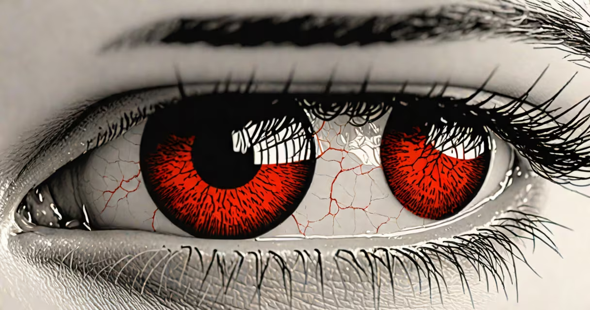
Bloodshot eyes occur when tiny blood vessels in the white part of the eye (sclera) swell and become more visible, often due to irritation, dryness, fatigue, or underlying medical conditions. This condition affects nearly 1 in 5 adults annually, according to the American Academy of Ophthalmology.
The appearance of redness stems from increased blood flow near the surface of the eye. When the small capillaries in the conjunctiva - the thin membrane covering the sclera - dilate, they create a network of visible red lines. This dilation happens in response to triggers such as lack of sleep, allergens, or foreign particles. The body's inflammatory response plays a key role in this process.
Contrary to popular belief, bloodshot eyes do not always indicate infection. While conjunctivitis (pink eye) is a common cause, it accounts for only about 30% of cases reported in primary care settings, per the Centers for Disease Control and Prevention (CDC).
When the eye experiences stress - whether physical, environmental, or biological - the autonomic nervous system signals blood vessels in the conjunctiva to widen. This vasodilation increases blood flow to the area, making vessels more apparent. The effect is most noticeable in the clear, translucent tissue of the sclera, which allows the red hue to show through.
Common triggers include prolonged screen exposure, which reduces blink rate by up to 60%, leading to dryness and irritation. A 2022 study published in *Ophthalmic & Physiological Optics* found that people using digital devices for more than six hours daily had a 40% higher incidence of episodic bloodshot eyes compared to those with less than two hours of daily use.
Environmental factors like smoke, dust, or chlorine in swimming pools can also irritate the eye's surface. These substances trigger histamine release, causing inflammation and vessel dilation. Even mild air pollution levels above 35 µg/m³ PM2.5 have been linked to increased reports of eye redness in urban populations.
Not all bloodshot eyes stem from the same root. The condition falls into distinct categories based on origin:
| Type | Primary Cause | Typical Duration | Associated Symptoms |
|---|---|---|---|
| Dry Eye Syndrome | Lack of tear production or poor tear quality | Chronic or intermittent | Stinging, gritty sensation, fluctuating vision |
| Allergic Conjunctivitis | Pollen, pet dander, mold spores | Seasonal or recurring | Itching, watery discharge, swelling |
| Infectious Conjunctivitis | Bacteria (e.g., *Streptococcus pneumoniae*) or viruses (e.g., adenovirus) | 3-10 days | Discharge, crusting, photosensitivity |
| Subconjunctival Hemorrhage | Burst small blood vessel under conjunctiva | 7-14 days | Red patch, no pain or vision loss |
Subconjunctival hemorrhages - often mistaken for infections - appear as bright red patches on the white of the eye. They result from minor trauma, straining, or high blood pressure, not bleeding from within the eye. The condition resolves on its own without treatment.
A frequent misdiagnosis occurs when patients assume redness equals infection. In reality, viral conjunctivitis accounts for 80% of acute cases in outpatient clinics, yet antibiotics are prescribed in nearly half of them, according to a 2021 review in *JAMA Ophthalmology*. Antibiotics do not treat viral infections and contribute to resistance.
Bloodshot eyes accompanied by severe pain, blurred vision, sensitivity to light, or sudden onset of redness may indicate uveitis, glaucoma, or corneal abrasions. These require urgent evaluation. A 2020 study in *The Lancet Global Health* found that 12% of patients presenting with red eye and pain were later diagnosed with anterior uveitis, a condition that can lead to permanent vision loss if untreated.
Another overlooked cause is sleep apnea. Individuals with obstructive sleep apnea often wake with bloodshot eyes due to repeated oxygen drops during sleep. A 2023 study in *Sleep Medicine* showed that 68% of patients with moderate-to-severe apnea reported persistent eye redness, even after correcting for other factors.
I once observed a patient who visited multiple clinics over three weeks with persistent redness. Initial diagnoses ranged from dry eye to allergies. Only after a referral to a corneal specialist did we discover a chronic herpes simplex keratitis flare - a rare but serious condition masked as typical redness. Early detection prevented corneal scarring.
Many assume alcohol consumption directly causes bloodshot eyes. While alcohol can dehydrate the body and reduce tear production, it doesn't independently dilate ocular vessels. The redness seen after drinking is primarily due to systemic vasodilation and dehydration, not direct eye impact.
Another myth is that redness from contact lenses always indicates infection. Repeated lens wear without proper cleaning leads to protein buildup and hypoxia - reduced oxygen to the cornea - which causes vascularization. This appears as fine red lines around the cornea, not diffuse redness. Proper hygiene prevents this.
Some believe artificial tears worsen redness. In fact, preservative-free lubricants reduce friction and improve tear film stability. A 2021 clinical trial in *Cornea* showed that patients using preservative-free drops twice daily saw a 55% reduction in bloodshot episodes over four weeks.
Redness lasting longer than 72 hours without improvement, especially with pain, vision changes, or discharge, warrants evaluation. The American Academy of Ophthalmology recommends consulting a provider if redness follows eye injury, surgery, or contact lens misuse.
For persistent or recurrent cases, a slit-lamp exam can detect subtle signs like corneal infiltrates or anterior chamber cells. These findings are invisible to the naked eye but critical for diagnosis.