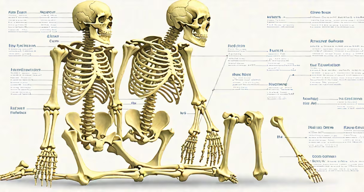
A full human skeleton is the complete set of 206 bones organized in their natural anatomical configuration, forming the rigid structure that supports the body, protects important organs, and enables movement. It includes the axial skeleton (skull, vertebral column, rib cage) and the appendicular skeleton (limbs, shoulder girdle, pelvic girdle).
The structure is derived from cadaver dissection and standardized anatomical models. Modern representations often use synthetic materials like plastic or resin to replicate bone density and joint articulation. These models are used in medical education, forensic science, and physical therapy training.
According to the National Library of Medicine, the human skeleton contains 206 bones in adults, though this number varies slightly during development due to fusion of bones such as the sacrum (five vertebrae fusing into one) and the coccyx (three to four fused bones).
Bones are living tissue composed primarily of collagen and calcium phosphate. They grow, remodel, and repair through cellular activity regulated by hormones like parathyroid hormone and calcitonin. The skeleton functions as a mechanical scaffold: muscles attach via tendons to bones, generating motion at joints.
For example, the femur (thigh bone) connects to the pelvis at the hip joint, allowing flexion, extension, and rotation. The knee joint-a hinge joint-permits bending and straightening of the leg. These movements are coordinated by the nervous system and supported by ligaments stabilizing the joints.
Internal cavities within bones house red and yellow marrow. Red marrow produces blood cells; yellow marrow stores fat. The skull encloses the brain, while the rib cage shields the heart and lungs. The spine protects the spinal cord, which transmits neural signals between the brain and body.
The full human skeleton consists of two major divisions:
| Skeletal Division | Number of Bones | Key Functions |
|---|---|---|
| Axial Skeleton | 80 | Supports head, neck, trunk; protects brain, spinal cord, heart, lungs |
| Appendicular Skeleton | 126 | Enables locomotion and manipulation; includes limbs and girdles |
The axial skeleton includes the skull (22 bones), hyoid bone (1), vertebral column (26), and thoracic cage (25). The appendicular skeleton comprises the pectoral girdle (4), upper limbs (64), pelvic girdle (2), and lower limbs (60).
Each bone has a specific name and function. For instance, the sternum (breastbone) forms the front of the rib cage and anchors the ribs. The scapula (shoulder blade) provides attachment points for muscles involved in arm movement.
Many assume the skeleton is static, but it's metabolically active. Bone remodeling occurs continuously-old bone is resorbed by osteoclasts and new bone formed by osteoblasts. This process maintains calcium balance and repairs microdamage from daily stress.
Another misconception is that skeletons are always symmetrical. While bilateral symmetry is typical, asymmetry can occur due to injury, disease, or congenital conditions. For example, scoliosis causes lateral curvature of the spine, altering the normal alignment of the vertebral column.
Some believe skeletons are fragile. In reality, adult human bone can withstand compressive forces exceeding 10,000 psi-roughly equivalent to a car pressing down on a single femur. This strength comes from the combination of organic matrix and mineralized crystals.
Medical students use full human skeletons to learn spatial relationships. A cadaver-based lab session at Johns Hopkins University requires students to identify 150+ bones in under three hours, with accuracy measured against standard anatomical atlases.
Forensic anthropologists analyze skeletal remains to estimate age, sex, stature, and cause of death. In one case documented by the American Academy of Forensic Sciences, a skeleton found in a forest was identified as male, aged 35-45, with a healed fracture in the left tibia consistent with a fall from height. The victim's identity was confirmed using dental records.
Physical therapists use full human skeleton models to demonstrate joint mechanics. For example, understanding how the patella (kneecap) tracks within the femoral groove helps diagnose and treat chondromalacia patellae, a condition affecting cartilage under the kneecap.
I once used a full human skeleton model during a workshop at a community health fair. A child asked why the spine curved. I demonstrated how the lumbar vertebrae arch forward, creating a natural curve that absorbs shock. She then pointed out the tailbone-"That's where the tail goes?"-showing how even basic models spark curiosity.
The full human skeleton is not just a collection of bones-it's a changing, living structure essential to human form and function. Understanding its composition, arrangement, and role in physiology is foundational to medicine, biology, and personal health.