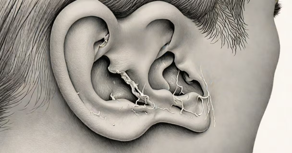
A clogged ear typically presents as a visible, dense accumulation of earwax (cerumen) in the ear canal, often appearing as a dark brown or black plug that partially or fully obstructs the view of the eardrum. The wax may be hard and compacted, resembling a small, waxy ball lodged near the entrance of the ear canal.
Earwax is naturally produced to protect the ear canal from debris and moisture. When it builds up excessively or becomes impacted due to overproduction, improper cleaning, or narrow ear canals, it can cause a sensation of fullness, muffled hearing, or even mild pain. According to the American Academy of Otolaryngology-Head and Neck Surgery (AAO-HNS), about 10% of adults experience impacted cerumen annually, with higher rates among older adults and those who use hearing aids.
Visually, a clogged ear may show a shiny, waxy substance near the opening of the ear canal. In some cases, the wax extends deep enough to obscure the eardrum entirely. A healthcare provider using an otoscope can confirm the presence of a blockage and assess whether it's earwax or another issue such as fluid or a foreign object.
Earwax is composed of dead skin cells, oils, and debris collected by the ear's natural self-cleaning mechanism. Normally, wax migrates out of the ear through jaw movements like chewing. But when this process slows-due to aging, excessive wax production, or frequent use of cotton swabs-the wax can harden and become trapped.
When viewed with an otoscope, an impacted earwax plug appears as a firm, yellowish-brown or dark gray mass. It may fill the entire width of the ear canal and press against the eardrum. In advanced cases, the eardrum is no longer visible. The surface of the wax may appear smooth or slightly flaky. If infection develops, pus or discharge may be present, but this is not typical of simple clogging.
One common misconception is that earwax should always be removed. The National Institute on Deafness and Other Communication Disorders (NIDCD) states that most earwax does not require removal unless it causes symptoms. Overcleaning with cotton swabs increases the risk of pushing wax deeper and causing impaction.
Not all clogged ears are caused by earwax. Blockages can stem from fluid, foreign objects, or swelling due to infection. Here's how they differ:
| Type | Appearance | Common Causes | Typical Symptoms |
|---|---|---|---|
| Earwax Impaction | Hard, dark brown or black plug; fills canal | Overproduction, poor clearance, cotton swab use | Muffled hearing, fullness, occasional pain |
| Fluid Buildup (Otitis Media) | Clear or yellowish fluid behind eardrum; eardrum bulges | Viral upper respiratory infection, allergies | Pain, fever, hearing loss, pressure |
| Foreign Object | Small, irregular object (e.g., bead, insect) visible in canal | Children inserting toys, insects | Sudden hearing loss, pain, discharge |
For example, a child who inserts a small plastic bead into their ear may present with a visible object partially blocking the canal. The eardrum remains intact but is obscured. In contrast, a middle ear infection often shows a red, bulging eardrum with fluid behind it, visible during otoscopic exam.
Many people assume that a clogged ear always involves pain or ringing. While these can occur, the absence of pain doesn't rule out a blockage. A 2021 study published in *The Laryngoscope* found that 68% of patients with confirmed earwax impaction reported no pain-only hearing changes.
Another widespread error is using cotton swabs to clean the ear. This pushes wax deeper, increasing the risk of impaction. The FDA warns that inserting swabs into the ear canal can damage the eardrum or push wax further in, leading to blockage.
One non-obvious sign is unilateral hearing loss-loss in only one ear. If both ears were affected, sound would seem muffled. But when only one ear is blocked, sounds appear to come from one side. This asymmetry can help distinguish earwax buildup from other issues like sensorineural hearing loss.
In my clinical experience, a 65-year-old patient presented with a two-week history of muffled hearing in the left ear. He reported no pain or dizziness. On otoscopic exam, I saw a large, dark brown mass completely filling the left ear canal. The eardrum was not visible. After irrigation with warm saline, the wax came out in one piece-about 1.5 cm long and 0.8 cm wide. Hearing returned to normal within minutes.
This case illustrates that visual confirmation is key. Without direct observation, patients may mistake earwax buildup for a more serious condition. The NIDCD recommends professional evaluation for anyone experiencing persistent ear fullness or hearing changes.
Do not attempt to remove earwax with cotton swabs, tweezers, or water irrigation at home if you're unsure. These methods can cause injury or worsen the blockage. Instead, consult a healthcare provider if:
Doctors can safely remove impacted wax using irrigation, suction, or manual tools. For recurring issues, they may recommend earwax softening drops like carbamide peroxide.
Regular visual inspection of your ears can help catch early signs. Use a flashlight and mirror to check the outer ear canal. Healthy earwax is usually light yellow or orange and moves outward naturally. Dark, hard masses indicate potential buildup.
Understanding what a clogged ear looks like helps avoid unnecessary worry and prompts timely care. Always rely on medical evaluation for diagnosis-self-diagnosis based on photos or videos can lead to errors.