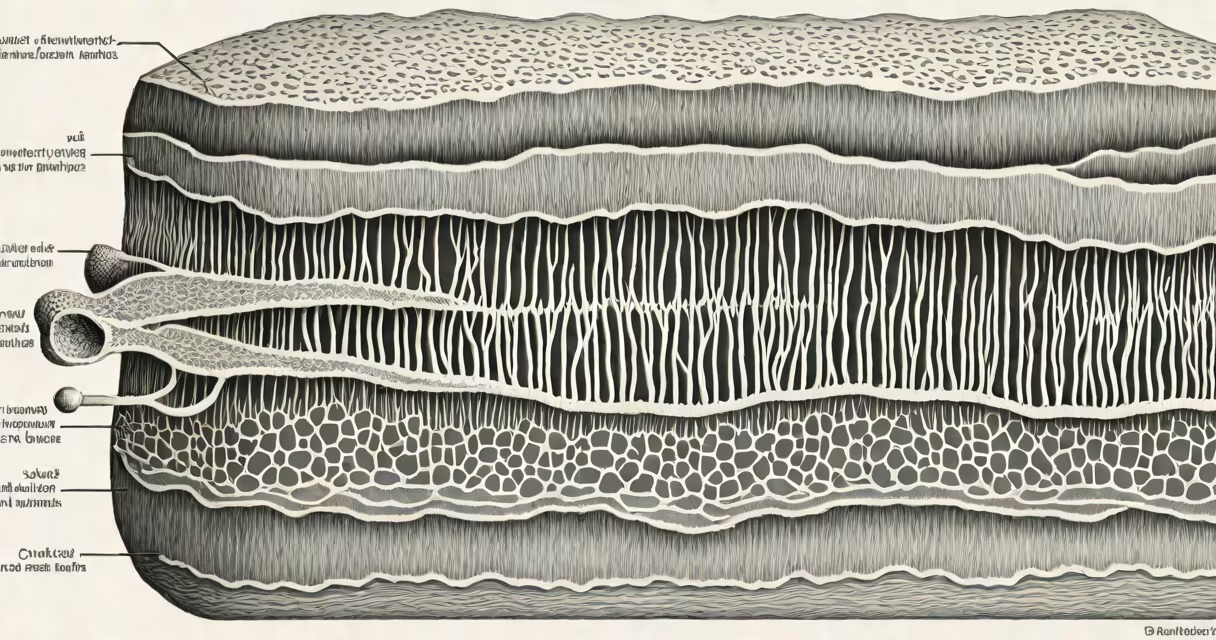
Smooth muscle cells generate sustained, slow contractions to regulate involuntary functions in internal organs such as the digestive tract, blood vessels, and bladder. These cells operate without conscious control and are essential for maintaining homeostasis.
Unlike skeletal muscle, which contracts rapidly and voluntarily, smooth muscle cells respond to signals from the autonomic nervous system and local chemical changes. Their primary role is to modulate flow-of blood, food, or fluids-through hollow organs by contracting and relaxing in coordinated waves.
These cells are found in the walls of the stomach, intestines, uterus, urinary bladder, and arteries. They enable processes like peristalsis (wave-like motion pushing food through the gut) and vasoconstriction (narrowing blood vessels to regulate blood pressure).
Smooth muscle cells contract through a process called excitation-contraction coupling. When stimulated by neurotransmitters, hormones, or stretch, calcium ions enter the cell and bind to calmodulin. This complex activates myosin light-chain kinase, which phosphorylates myosin, allowing it to interact with actin filaments and generate force.
Unlike skeletal muscle, smooth muscle lacks sarcomeres-the repeating units of actin and myosin seen in striated muscle. Instead, filaments are arranged in a web-like pattern, enabling contraction in multiple directions. This allows smooth muscle to squeeze, stretch, and reshape organ walls efficiently.
A key feature is their ability to sustain contraction for long periods without fatigue. A single contraction can last seconds to minutes, ideal for maintaining tone in blood vessel walls or keeping the bladder closed until voiding.
Three main types exist based on structure and location:
| Type | Structure | Location | Function |
|---|---|---|---|
| Single-unit | Spindle-shaped; connected by gap junctions | Intestines, uterus, small blood vessels | Syncytial contraction; acts as a unit |
| Multilayered | Individual cells; no direct connections | Larger arteries, iris of eye | Independent control per cell |
| Visceral | Arranged in sheets; responsive to stretch | Stomach, bladder | Adapts to volume changes |
Single-unit smooth muscle, common in the intestines, contracts as a group due to gap junctions that allow electrical signals to pass between cells. This enables synchronized peristaltic waves that move contents forward.
Arteries larger than 100 micrometers often contain multilayered smooth muscle. Each cell responds independently to neural input, allowing fine-tuned regulation of blood flow. For example, during stress, sympathetic nerves trigger constriction in renal arteries to redirect blood to muscles.
Many assume smooth muscle cells are "weaker" than skeletal muscle because they contract slowly. But their strength lies in endurance and precision. A single smooth muscle cell can generate enough force to close a blood vessel diameter of 2 mm.
Another misconception is that smooth muscle is inactive unless stimulated. In reality, many smooth muscle tissues exhibit spontaneous activity. The intestinal wall, for instance, generates baseline contractions even without nerve input-this is known as intrinsic tone.
Smooth muscle is not controlled by motor neurons in the same way as skeletal muscle. Instead, it receives input from autonomic nerves (sympathetic and parasympathetic systems), hormones (like adrenaline), and local factors (such as oxygen levels or pH).
Consider the arterioles-small branches of arteries that regulate blood flow into capillary beds. When blood pressure drops, baroreceptors in the carotid sinus detect the change and send signals via the glossopharyngeal nerve to the brainstem.
The medulla oblongata then increases sympathetic output. Norepinephrine released from nerve endings binds to alpha-1 receptors on smooth muscle cells in arterioles. This triggers calcium influx, leading to contraction and vasoconstriction.
As vessel diameter decreases from 200 μm to 100 μm, resistance increases fourfold (per Poiseuille's law), raising blood pressure. This process stabilizes circulation within seconds-without any conscious effort.
I've observed this mechanism firsthand during a physiology lab using isolated rat mesenteric arteries. When exposed to norepinephrine, the vessel contracted visibly under a microscope. After washing out the drug, relaxation occurred gradually over 2-3 minutes-consistent with smooth muscle's slow kinetics.
Smooth muscle differs from skeletal and cardiac muscle in several ways:
Despite these differences, all three types rely on actin and myosin interaction. The variation lies in regulatory mechanisms and structural organization.
Dysfunction in smooth muscle contributes to major health conditions. Hypertension arises when vascular smooth muscle remains overly constricted. Gastrointestinal disorders like irritable bowel syndrome involve abnormal contractions in intestinal smooth muscle.
Drugs targeting smooth muscle include calcium channel blockers (used in hypertension) and beta-agonists (used in asthma to relax bronchial smooth muscle). These medications work by interfering with the signaling pathways that activate contraction.
According to the National Institutes of Health (NIH), smooth muscle accounts for about 60% of total body mass in the cardiovascular system alone. The American Heart Association notes that smooth muscle dysfunction is central to atherosclerosis, where chronic inflammation leads to stiffening and reduced contractility of arterial walls.
Understanding what smooth muscle cells do helps explain how everyday bodily functions-from digesting lunch to regulating blood pressure-occur automatically. Without them, important systems would fail within minutes.
Smooth muscle cells perform critical, silent work throughout the body. They manage internal flows, maintain pressure, and support organ function-all without requiring your attention.